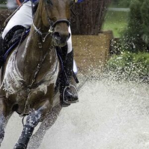
LAMENESS AND POOR PERFORMANCE
It has been reported that about 90% of poor performance cases can be attributed to lameness, either clinical (obvious lameness) or sub-clinical (lameness not readily visible under normal exam conditions).
It is logical that noticeable lameness causes horses to perform below their potential, but sub-clinical lameness can be an even greater problem. Clinical lameness can be quickly recognized, investigated, and corrected. In horses with sub-clinical lameness, however, the disease process remains undetected and untreated. It is allowed to progress, resulting in irreversible damage to the structure of joints, secondary lameness, muscle pain, behaviour problems, impaired performance and economic losses.
Early diagnosis and intervention can stop minor problems from deteriorating, preserving long term soundness and maximizing performance.
Most of my clients present every horse in their stable, on a regular basis, for physical exams. This enables the identification of subtle or sub-clinical problems.
Clinical Lameness
A horse is clinically lame if it has a visible limp or asymmetric gait. It will try to lift its weight off the sore leg and place more weight on the sound legs. A “head-nod” results. (When the sore front leg hits the ground, the horse lifts its head up to shift weight to the back legs and off the sore front leg. When the sound front leg hits the ground, the head nods down, loading that leg excessively.) Sometimes, when a horse is very lame in a hind leg, the horse will nod its head down to shift weight onto the front legs and off of the hind legs. Sometimes, a horse with a sore hind leg will lift its pelvis higher on the lame side (called a hip-hike).
Lameness is only visible (clinical) when one leg is relatively more painful than the opposite leg. Both legs can be sore, but as long as the pain is unequal, the horse will protect the more sore side and the head nod will be evident. There are various degrees of clinical lameness ranging from an inconsistent or almost imperceptible limp to an inability to bear any weight at all on the affected leg.
Sub-Clinical Lameness
Sub-clinical lameness is lameness that you can not see under normal conditions. Bilateral lameness, lameness in all four legs, and lameness that only manifests under extreme stress or speed is sub-clinical.
Bilateral lameness is often inapparent. If a horse’s legs are equally sore, he will not favor one and will not limp. Instead, he will shorten his stride, develop back or muscle pain, perform below expectations, make breaks, “stop” in the last part of a race, refuse jumps, make mistakes of stride in dressage tests, tie up, blow after working, have a longer than normal recovery, or develop behaviour problems such as pulling, bucking, and rearing. Many horses just develop a poor attitude to work. “Bleeding” or Exercise Induced Pulmonary Hemorrhage and dorsal displacement of the soft palate (“flipping the palate”) are common presenting complaints.
Some lameness only shows up at high speed or under extreme stress such as in the last part of a race. Some will manifest only with a rider or doing particular movements like flying changes or lateral work. Some will appear on a lunge line or on particularly hard, soft, or irregular or unstable footing. Some only present in the cart and not in-hand. Once again, these lameness cases are often presented for performance and behavior problems, back, or other muscle pain.
Lameness In My Practice
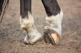
In my practice, the majority of horses presented for lameness or performance problems have one or more of the following: 1) foot pain including sole bruises, abscesses and, corns; 2) arthritis (joint inflammation); 3) tendonitis (a bowed tendon) or; 4) suspensory desmitis. Bowed tendons and suspensory desmitis present as clinical lameness and there is obvious pain, heat, and swelling. By far, the most common sub-clinical lameness or performance problems involve joint and foot pain. In many cases these conditions are both present.
Arthritis
Arthritis is a term that means “joint inflammation” (arth-joint, itis – inflammation). Inflammation occurs in joints when they are placed under stress in excess of what they have adapted for. This stress can be sudden and severe (stepping in a hole, taking a bad step on poor footing, or some other accident), or it can be repetitive and low grade (wear and tear).
Horses are designed for eating grass and running away from the occasional predator. They are designed to land flat on their feet, load bones and joints evenly from side to side, and break over the middle of their toes. Unfortunately, not many horses have perfect conformation, perfect hoof balance, or work on perfect footing so stress is not distributed evenly. They are not born readily adapted for repeatedly pulling a sulky or carrying a rider around a track at top speed or over jumps. The idea behind training is to gradually increase the stress on a horse causing them to adapt to the work we expect them to do. In short, training a young horse or training a more mature horse down to race after a spell is constantly placing their joints under stress they have not adapted to. Therefore, inflammation occurs on an ongoing basis in most horses in training.
Joints are made up of the ends of two or more bones which are covered with cartilage and joined together by the joint capsule. The joint capsule is lined by the synovial membrane. This membrane is very important as it produces the synovial fluid (joint fluid) that lubricates, protects and nourishes the joint cartilage. In a healthy joint, synovial fluid is thick like syrup. It is replaced every 24 hours or so on an ongoing basis.
Inflammation in joints begins with synovitis and capsulitis. In synovitis and capsulitis, enzymes are produced that breakdown joint fluid, making it thin and watery. It no longer lubricates and protects the joint properly. With a lack of nourishment and lubrication, the cartilage surface of the joint becomes abraded. Over a more extended period of time, the sub-chondral bone (bone underneath the cartilage in the joint) begins to change.
Over time, then, synovitis and capsulitis will progress to sub-chondral bone disease and osteoarthritis. This entire process is referred to as arthritis or degenerative joint disease (DJD). X-rays only show bone, so relatively advanced DJD is the first stage that is reliably visible on radiographs.
It is much better to identify and treat joint problems before they are visible on radiograph. If inflammation is stopped, the synovial membrane will make new fluid that will remain thick and sticky. If the breakdown of synovial fluid is the only damage that has occurred, a completely normal joint environment will be restored. If the cartilage surface has been damaged, some treatments can provide repair, and a normal joint can be created. Once bone has changed, however, it cannot be reversed. Thick, healthy joint fluid will stop rough bones from rubbing together in the joint, and DJD will be arrested, but a truly normal joint cannot be restored.
Treatment
Since the primary goal of therapy is to stop inflammation and to stop the progression of degenerative joint disease, the treatment of choice in most cases is intra-articular cortisone. Cortisones are very effective anti-inflammatories, and remain the treatment of choice in human medicine for intra-articular therapy.
There are several different types of cortisone that can be used in joints. Controlled studies have shown that all cortisones reduce inflammation and that most improve the health of joint cartilage. Triamcinolone (Vetalog, Kenalog, or Kenacort), Isoflupredone (Predef 2X), and Betamethasone (Celestone Soluspan) have all been shown to be safe or beneficial for joint cartilage. Methyl Prednisolone (Depo-Medrol or Vetacortyl) is likely safe in low doses, but can impair the healing of joint cartilage if given too frequently or in large amounts. Triamcinolone has been anecdotally linked to laminitis, but the relationship has not been confirmed. It has never been caused in healthy horses at normal doses and has not been caused experimentally using doses up to six times those commonly used.
NSAIDS like Bute and Banamine tend to kill pain better than they reduce inflammation in joints, so they are not a sufficient treatment in most cases.
Once inflammation is resolved and DJD is arrested, the second goal of therapy is to restore a normal joint environment. Hyaluronic acid (HA) is a building block for thick joint fluid, so supplementation may be useful. HA can be administered directly into a joint, however, it does not work very well if there is a great deal of inflammation present and it is generally used in joints along with cortisone. Studies have shown that IV HA (Hyonate or Legend) is as useful as intra-articular treatments, and recent research indicates that oral administration of HA may be helpful.
If cartilage damage has already occurred, then it can be beneficial to treat horses with a product that can stimulate joint repair or provide the building blocks for cartilage repair. Adequan, Glucosamine Sulphate, and Pentosan may be used for this purpose. Glucosamine Sulphate supplementation increases the body’s production of hyaluronic acid as well.
Additional medications are available to treat arthritis including some homeopathic treatments that reduce inflammation and stimulate joint healing. The homeopathic medications I mainly use are Traumeel and Zeel.
Finally, the third goal of therapy is to prevent reoccurrence of lameness. Adequan, Glucosamine, or Pentosan can be given regularly to reduce inflammation and repair cartilage. They can keep inflammation at bay in sound horses in training, and they can increase the interval between joint injections in horses with lameness problems. Optimal shoeing and good footing are of utmost importance, and adjustments to the training regimen may be helpful in some cases.
Summary
- The products of inflammation are enzymes that damage the joint.
- Early diagnosis and treatment will preserve normal joint structure and function maximizing long term soundness and performance.
- The first goal of therapy is to stop inflammation and, therefore, to stop the progression of degenerative joint disease.
- The second goal of therapy is to restore the most normal joint environment possible.
- The third goal of therapy is to prevent reoccurrence of the problem.3
Dr Corinne Hills, Pro-Dosa International Ltd., 34 Ryan Road, RD 4, Pukekohe, New Zealand.
Phone: +64 27 238 8482 Email: info@pro-dosa.com Website: www.pro-dosa.com FB: ProDosaBoost